Table of Contents
Comprehensive Introduction to 5-ALA/ICG Molecular Fluorescence Imaging Technology in Medical Endoscopy
Molecular fluorescence imaging is a revolutionary technology in the field of medical endoscopy in recent years, which achieves real-time and accurate visualization diagnosis and treatment through the specific binding of specific fluorescent markers (such as 5-ALA, ICG) to diseased tissues. The following provides a comprehensive analysis of technical principles, clinical applications, comparative advantages, representative products, and future trends.
1. Technical principles
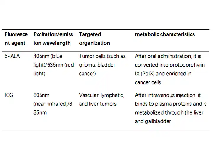
(1) The mechanism of action of fluorescent markers

(2) Composition of imaging system
Excitation light source: Specific wavelength LED or laser (such as blue light excitation of 5-ALA).
Optical filter: filters out interference light and only captures fluorescence signals.
Image processing: overlaying fluorescent signals with white light images (such as real-time fusion display of PINPOINT system).
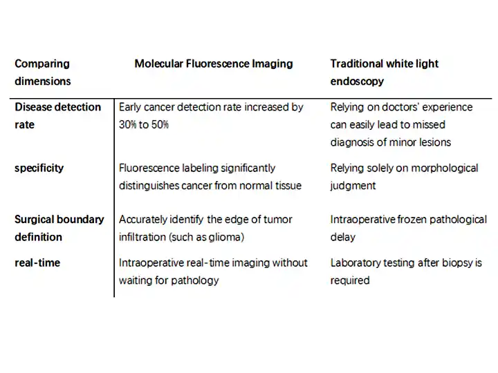
2. Core advantages (vs traditional white light endoscopy)

3. Clinical application scenarios
(1) 5-ALA fluorescence endoscope
Neurosurgery:
Glioma resection surgery: PpIX fluorescence labeling of tumor boundaries increases the total resection rate by 20% (if approved for use with GLIOLAN).
Urology:
O Diagnosis of bladder cancer: fluorescent cystoscopy (such as Karl Storz D-LIGHT C) reduces the recurrence rate.
(2) ICG fluorescence endoscope
Hepatobiliary Surgery:
Liver cancer resection surgery: precise resection of ICG retention positive areas (such as Olympus VISERA ELITE II).
Breast Surgery:
Sentinel lymph node biopsy: ICG tracing replaces radioactive isotopes.
(3) Multi modal joint application
Fluorescence+NBI: Olympus EVIS X1 combines narrowband imaging with ICG fluorescence to improve the diagnostic rate of gastric cancer.
Fluorescence+ultrasound: ICG labeling of pancreatic tumors guided by endoscopic ultrasonography (EUS).
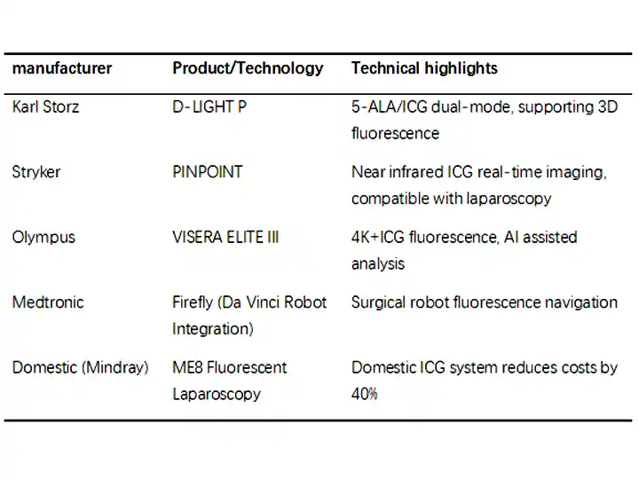
4. Representing manufacturers and products

5. Technical challenges and solutions
(1) Fluorescence signal attenuation
Problem: The duration of 5-ALA fluorescence is short (about 6 hours).
Solution:
O Intraoperative administration in batches (such as multiple perfusion during bladder cancer surgery).
(2) False positive/false negative
Problem: Inflammation or scar tissue may mistake fluorescence.
Solution:
Multispectral analysis (such as distinguishing PpIX from autofluorescence).
(3) Cost and Popularization
Problem: The price of fluorescent endoscopic systems is high (approximately 2 to 5 million yuan).
Breakthrough direction:
Domestic substitution (such as Mindray ME8 system).
Disposable fluorescent endoscope (such as Ambu aScope ICE).
6. Future Development Trends
(1)New fluorescent probe:Tumor specific antibody fluorescent labeling (such as EGFR targeted probes).
(2) AI quantitative analysis:Automated grading of fluorescence intensity (such as using ProSense software to assess tumor malignancy).
(3)Nanofluorescence technology:Quantum dot (QDs) labeling enables multi-target synchronous imaging.
(4) Portability:Handheld fluorescent endoscope (such as used for screening in primary hospitals).
summarize
Molecular fluorescence imaging technology is changing the paradigm of tumor diagnosis and treatment through "precise labeling+real-time navigation":
Diagnosis: The detection rate of early cancer has significantly increased, reducing unnecessary biopsies.
Treatment: The surgical margin is more precise, reducing the risk of recurrence.
Future: With the diversification of probes and integration of AI, it is expected to become a standard tool for "intraoperative pathology".
Copyright © 2025.Geekvalue All rights reserved.Technical Support:TiaoQingCMS