
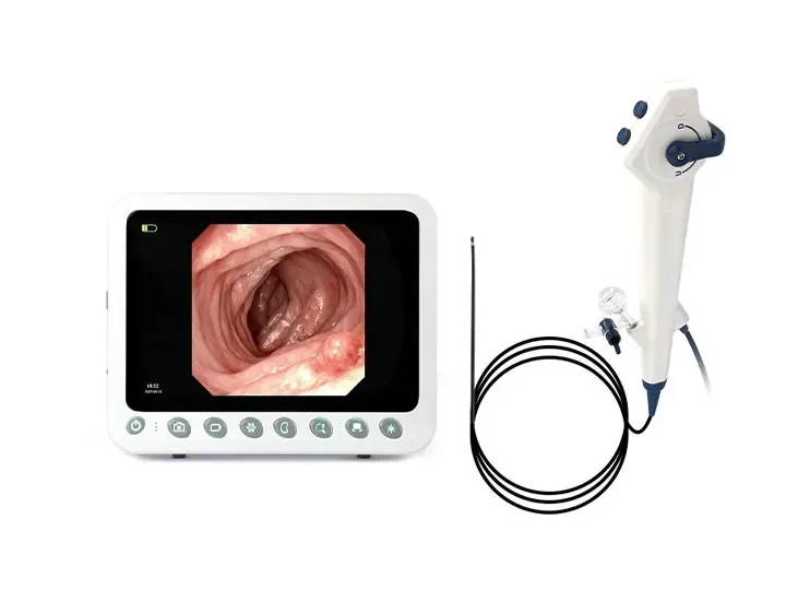
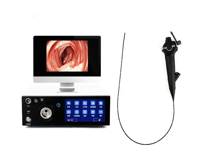
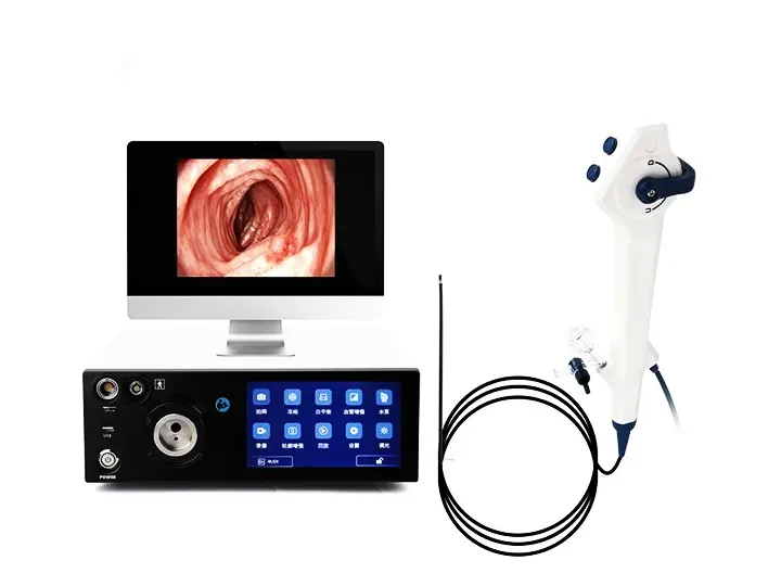
Wide Compatibility
Wide compatibility:Ureteroscope, Bronchoscope, Hysteroscope, Arthroscope, Cystoscope, Laryngoscope, Choledochoscope
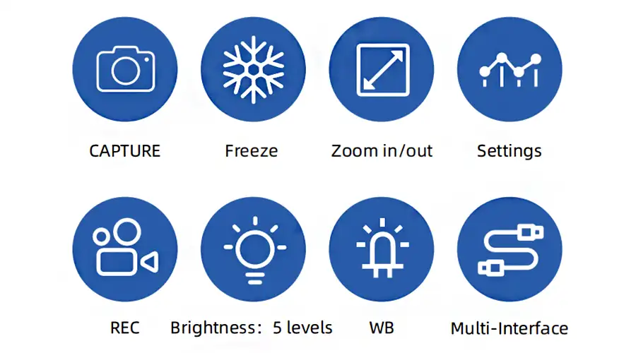
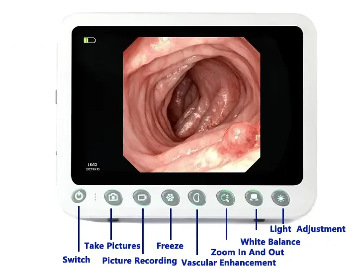
Capture
Freeze
Zoom In/Out
Image Settings
REC
Brightness: 5 levels
WB
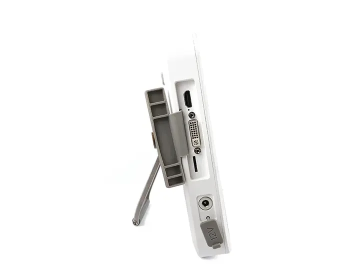
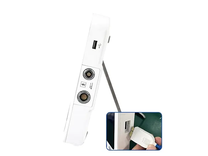
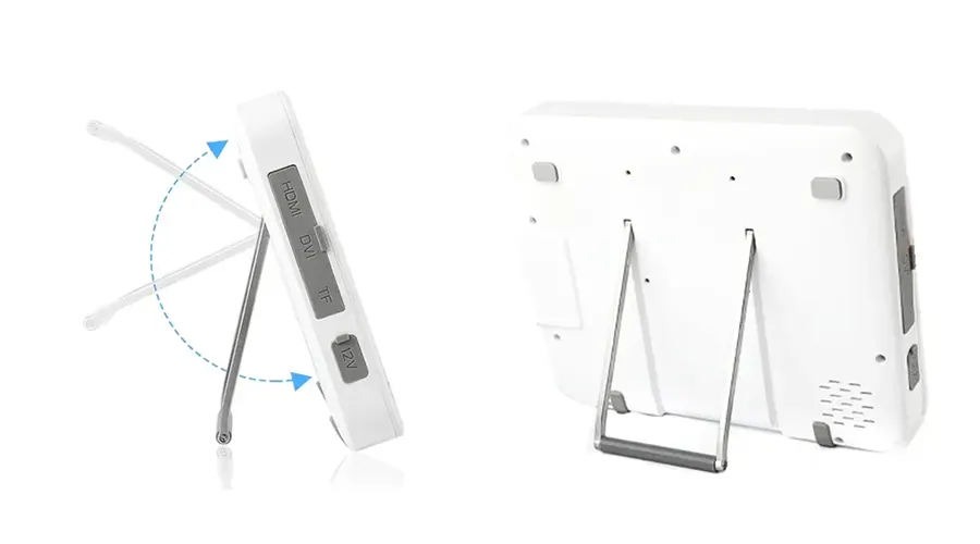
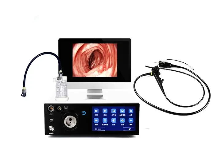
Multi-Interface
1280×800 Resolution Image Clarity
10.1" Medical Display,Resolution 1280×800,
Brightness 400+,High-definition
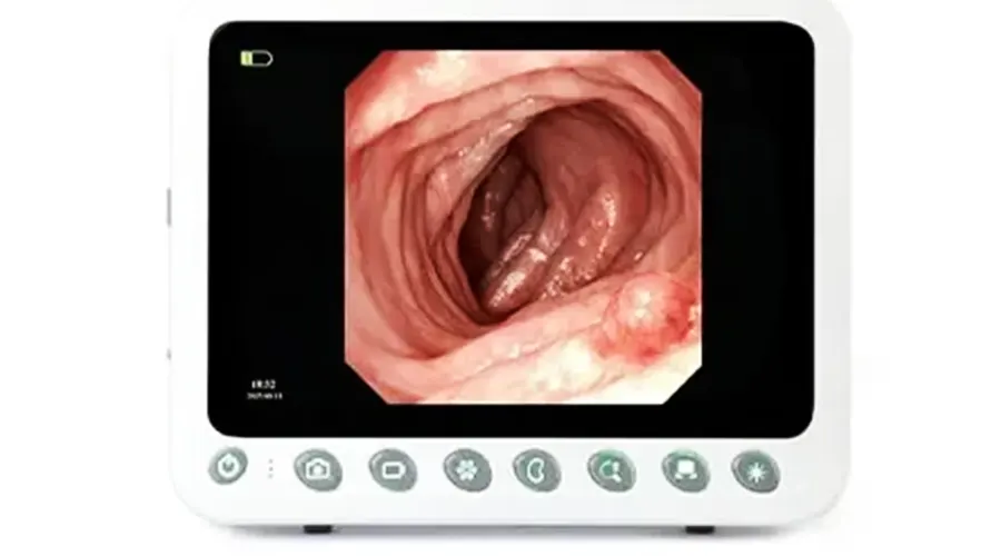
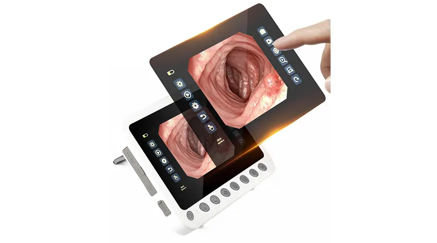
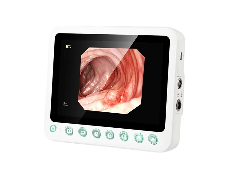
High-definition Touchscreen Physical Buttons
Ultra-responsive touch control
Comfortable viewing experience
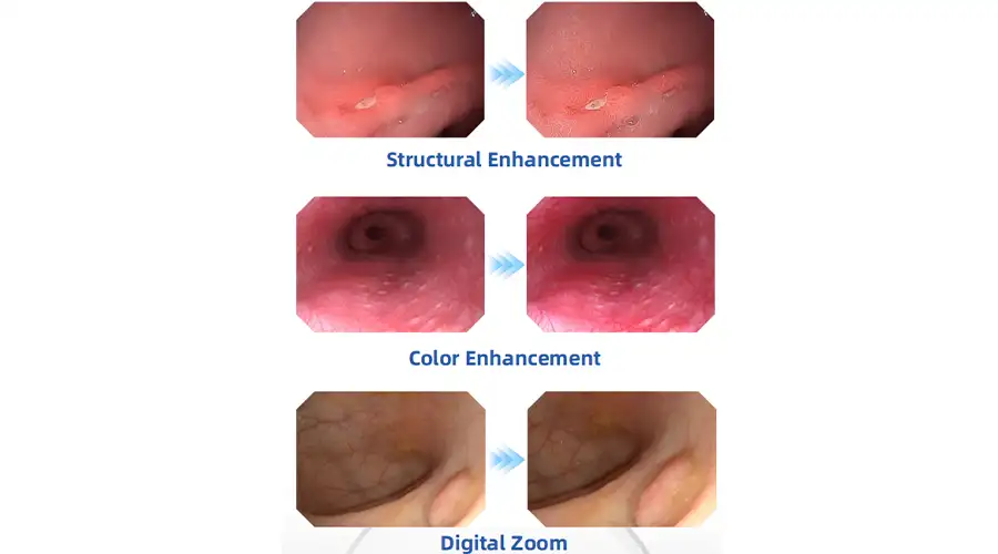
Clear Visualization For Confident Diagnosis
HD digital signal with structural enhancement
and color enhancement
Multi-layer image processing ensures every detail is visible
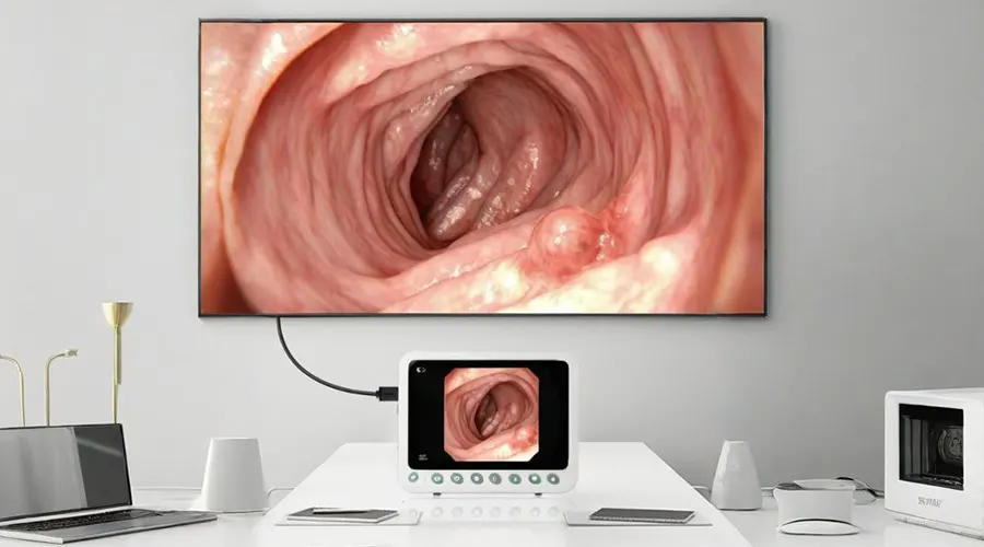
Dual-screen Display For Clearer Details
Connect via DVI/HDMI to external monitors - Synchronized
display between 10.1" screen and large monitor
Adjustable Tilt Mechanism
Slim and lightweight for flexible angle adjustment,
Adapts to various working postures (standing/sitting).
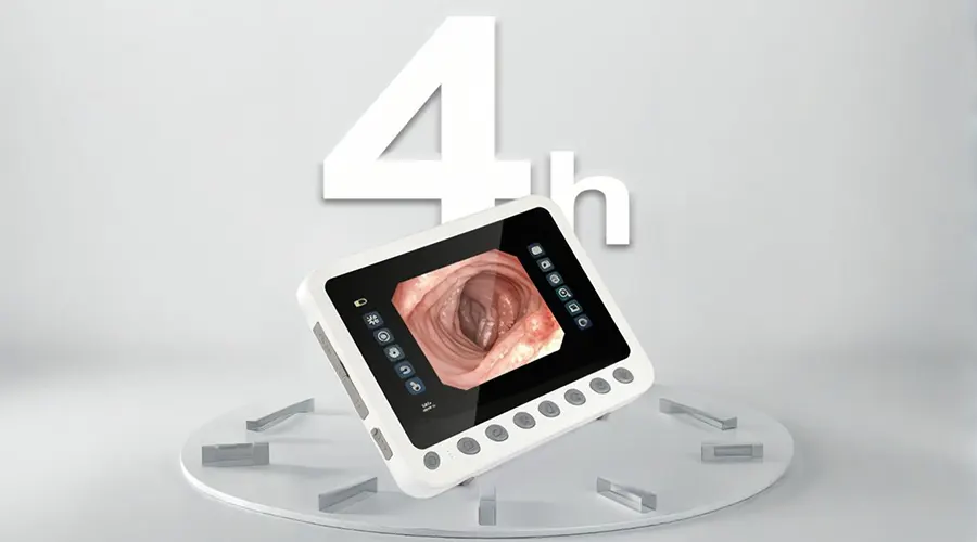
Extended Operation Time
Ideal for POC and ICU examinations - Provides
doctors with convenient and clear visualization
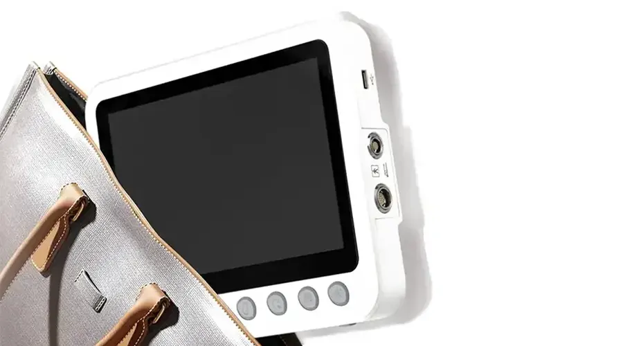
Portable Solution
Ideal for POC and ICU examinations - Provides
doctors with convenient and clear visualization


Urological Endoscopy is the gold standard for the diagnosis and treatment of urinary system diseases, achieving non-invasive exploration, accurate diagnosis and minimally invasive treatment through natural cavities or tiny incisions. The following is a comprehensive analysis from six dimensions:
1. Technical principles and equipment evolution
Core components
Optical system: 4K ultra-high-definition/3D imaging, NBI narrow-band light for early identification of tumors
Scope type:
▸ Hard scope (0°-70° viewing angle, used for bladder/ureter)
▸ Soft scope (270° bending, reaching the renal pelvis)
Working channel: supports laser fiber, stone basket, biopsy forceps and other instruments
Technology iteration
From fiberscope to electronic scope: pixel increase 100 times (now up to 500,000 pixels)
From white light to intelligent imaging: fluorescent markers (such as 5-ALA) make cancer cells self-luminous
2. Full spectrum of clinical applications
Disease field Diagnostic application Therapeutic application
Bladder Tumor staging, interstitial cystitis evaluation Tumor resection (TURBT), lithotripsy
Ureter Stricture positioning, foreign body detection Stent placement, laser lithotripsy
Kidney Hematuria tracing, space-occupying lesion biopsy Percutaneous nephrolithotomy (PCNL)
Prostate hyperplasia assessment and enucleation (HoLEP)
III. Comparison of mainstream devices
Type Diameter Advantages Classic scenarios
Cystoscopy 16-22Fr Large channel and multi-instrument collaboration Prostate resection
Ureteroscopy 7.5-9.9Fr Active bending 270° Laser powderization of renal pelvic stones
Percutaneous nephroscope 18-30Fr Direct establishment of renal channel Staghorn stone removal
Disposable electronic scope 6.5Fr Zero risk of cross infection Outpatient rapid examination
IV. Essentials of surgical procedures (taking ureteroscopic lithotripsy as an example)
Preoperative
Three-dimensional CT planning of stone location, general anesthesia
Intraoperative
Insert soft endoscope under the guidance of guidewire, and holmium laser eats away stones to <2mm
Place double J tube to prevent stenosis if necessary
Postoperative
Drink 2000ml of water on the same day, and remove the catheter in 3 days
V. Complication prevention and control
Bleeding: plasma bipolar electrocoagulation
Infection: preoperative urine culture + targeted antibiotics
Perforation: real-time pressure monitoring during surgery (<40cmH₂O)
VI. Five major breakthrough directions in the future
AI real-time pathology: automatic distinction between low-grade and high-grade urothelial carcinoma under the microscope
Microrobot: magnetically controlled capsule endoscope to screen early lesions
Virtual reality training: doctors simulate surgery on 3D reconstructed organs
Biodegradable stents: no need for secondary removal after surgery
Targeted photodynamic therapy: accurate elimination of in situ cancer cells
Industry value summary
Uroscopic technology enables urology to achieve:
🔹 Diagnosis upgrade: early tumor detection rate increased by 3 times
🔹 Treatment innovation: 90% of stone surgeries do not require surgery
🔹 Patient benefits: hospital stay shortened to 1-2 days
With the integration of single-port laparoscope and endoscope, the future will usher in a new era of scarless surgery.
Faq
-
Will the medical uroscope machine examination be very painful?
Surface anesthesia or intravenous sedation will be used during the examination, and most patients only feel slight discomfort. The examination time is short, and they can recover after a short rest after surgery.
-
What diseases can medical uroscope machine treat?
It can be used for the diagnosis and treatment of stones, tumors, prostate hyperplasia, etc., and can be directly crushed or excised with laser or electric cutting equipment.
-
What are the special requirements for disinfection of medical uroscope machines?
Special sterilizers should be used for high-temperature treatment, and the mirror body pipeline should be thoroughly rinsed to prevent biofilm residue and ensure sterility standards are met.
-
Do I need to be hospitalized after medical uroscope machine examination?
Ordinary examinations do not require hospitalization. If treatment such as lithotripsy or resection is performed, observation for 1-2 days is necessary to confirm that there is no bleeding or infection before discharge.
Latest articles
-
How XBX Cystoscope Supplier Ensures Quality and Precision for Hospital Procurement
Discover how the XBX Cystoscope Supplier provides hospitals with high-precision, OEM-ready endoscopy systems built for reliability, safety, and consistent imagi...
-
How XBX Bronchoscope Factory Delivers Reliable OEM Systems
Discover how the XBX Bronchoscope Factory ensures quality and reliability through advanced OEM manufacturing, optical precision, and strict quality control.
-
How XBX Laparoscope Minimizes Surgical Trauma in Abdominal Surgery
Discover how the XBX Laparoscope reduces surgical trauma through precision imaging, minimal incisions, and faster recovery in modern abdominal procedures.
-
How XBX Hysteroscope Detects and Removes Uterine Polyps
Discover how the XBX Hysteroscope enables precise detection and removal of uterine polyps, improving accuracy, safety, and comfort in women’s health care.
-
What Is an XBX Flexible Ureteroscope for Stone Removal?
Learn how the XBX flexible ureteroscope improves access, visibility, and efficiency in ureteral stone management with 4K imaging and ergonomic control.
Recommended products
-
 Endoscope Image Processor Portable Host
The Endoscope Image Processor Portable Host enhances minimally invasive procedures with high-quality
-
 Medical gastroscopy equipment
Medical gastroscopy equipment provides HD imaging for endoscopy medical endoscopes, enhancing diagno
-
 XBX Repeating ENT Endoscope Equipment
Reusable ENT Endoscopes are medical optical instruments designed for examination of the ears, nose,
-
 XBX Medical Repeating Bronchoscope
Reusable bronchoscope refers to a bronchoscope system that can be used multiple times after professi
