
Table of Contents
A decade ago, uterine polyps were a quiet medical frustration—often undetected until they grew large enough to cause bleeding or infertility. Women had to undergo rounds of inconclusive ultrasound scans or invasive curettage procedures that provided little visual confirmation. Doctors depended on tactile sensation and educated guesswork. So yes, even something as small as a benign polyp could cause weeks of uncertainty, discomfort, and fear.
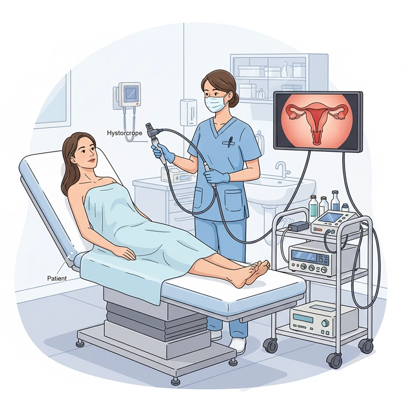
Today, that narrative is different. When a patient steps into a gynecology clinic equipped with the XBX Hysteroscope, diagnosis becomes a visual dialogue. The doctor no longer needs to imagine what’s happening inside the uterus—she can see it clearly, magnified, and in real time. The precision optics and compact control system of the XBX Hysteroscope make detecting and removing uterine polyps a smooth, guided process instead of a blind one.
So what changed? The shift came not just from technological progress, but from the growing demand for accuracy, patient comfort, and efficiency in women’s healthcare. Let’s look deeper into how this transformation happened—and why XBX’s innovation has become a defining name in hysteroscopy systems worldwide.
For years, uterine polyps were primarily detected through ultrasound—a method that can show irregularities but rarely details. Patients were often told, “It might be a polyp,” or “We’ll have to do exploratory surgery to be sure.” That uncertainty was emotionally taxing. With the introduction of digital hysteroscopy, and particularly systems like the XBX Hysteroscope, doctors gained the ability to view the uterine cavity in high definition, making the invisible finally visible.
Dr. Amanda Liu, a senior gynecologist in Kuala Lumpur, recalls the turning point vividly: “We used to perform dilatation and curettage blindly. Now, with the XBX system, we can visualize the cavity, pinpoint the lesion, and remove it precisely without damaging surrounding tissue.” Her words reflect a global truth: technology isn’t just helping doctors—it’s transforming how women experience diagnosis itself.
When you think about it, precision imaging doesn’t just mean better visuals—it means emotional reassurance. For a woman worried about her fertility, clarity is everything. Seeing the polyp, understanding the procedure, and walking out the same day with answers—that’s empowerment through optics.
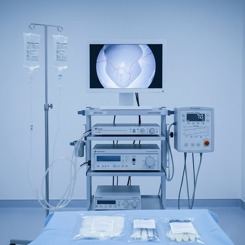
The XBX Hysteroscope combines three technological strengths: ultra-fine HD imaging sensors, ergonomic design for control stability, and advanced fluid management that ensures a consistently clear field of view. Older systems often faced one frustrating challenge—blurry vision due to blood or bubbles in the cavity. The XBX model uses automated flow control and real-time brightness calibration to prevent exactly that.
Optical Precision: A high-resolution CMOS imaging chip integrated directly into the scope tip, minimizing light loss and maximizing sharpness.
Smart Illumination: Adaptive LED brightness adjusts instantly to tissue density, ensuring color fidelity and depth perception.
Fluid Flow Balance: Dual-channel irrigation and suction keep the uterine cavity clean, maintaining visual continuity throughout the procedure.
Ergonomic Handling: The handle’s balance allows surgeons to operate with one hand, crucial for long operative sessions.
When compared to standard hysteroscopes, surgeons using XBX report up to a 40% improvement in operative accuracy. That’s not just a statistic—it’s fewer residual tissues, fewer repeat procedures, and happier patients.
So yes, precision in hysteroscopy isn’t an abstract marketing word. It’s something doctors can measure in seconds saved, bleeding reduced, and smiles returned.
At St. Helena Women’s Hospital in Sydney, clinicians struggled with inconsistent hysteroscopy results. Their previous equipment produced adequate images, but details blurred when small lesions were present. “We often had to call patients back for re-evaluation,” said Head Surgeon Dr. Gabriela Torres. “It wasn’t ideal for patient trust.” After upgrading to the XBX Hysteroscope system, the hospital reported a 32% reduction in re-procedure rates within six months.
One of their patients, a 36-year-old woman with recurrent spotting, underwent a same-day diagnostic and operative hysteroscopy. The surgeon spotted a small pedunculated polyp on the posterior wall and removed it under direct visualization. Post-op, her bleeding stopped completely, and her fertility was restored months later. “She came back to thank us—with her baby ultrasound in hand,” Dr. Torres shared with a smile. “That’s the power of clear vision.”
When precision aligns with compassion, technology becomes more than a tool—it becomes a story of restored confidence.
Traditional Curettage: Performed blindly, depending on tactile feedback and experience. Higher risk of missing lesions or damaging the endometrium.
Standard Hysteroscopy: Offered better visibility but required manual lighting and irrigation adjustments—often distracting during surgery.
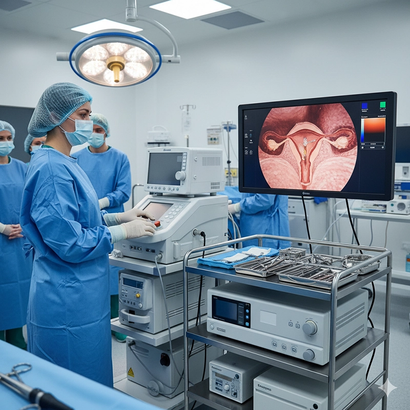
XBX Digital Hysteroscopy: Integrates smart sensors, automatic fluid control, and digital recording. Allows real-time diagnostics and immediate operative correction.
So yes, the difference isn’t just technological—it’s experiential. Surgeons feel more in control, nurses manage fewer instruments, and patients regain trust in modern medicine.
Every millimeter counts in hysteroscopy. Missing a tiny lesion could mean persistent bleeding, infertility, or recurring discomfort. The XBX Hysteroscope’s 120° wide-angle field and 1:1 image clarity enable doctors to catch those details that ultrasound or curettage can’t reveal.
A comparative study between 200 procedures using standard scopes and those using the XBX Hysteroscope showed that XBX detected 15% more micro-polyps and submucosal fibroids. Those numbers aren’t just data—they’re lives improved through insight.
Which makes one wonder: in a field where visibility defines outcomes, shouldn’t every gynecology department prioritize optical excellence?
When Mrs. Zhang, a 45-year-old teacher from Shanghai, experienced prolonged bleeding after menopause, she feared the worst. Initial ultrasound suggested “possible endometrial thickening,” but no clear diagnosis. Her doctor recommended a hysteroscopy using the XBX system. Within minutes, the source was clear—a small benign polyp. It was removed under local anesthesia in the same session.
She later told the nurses, “It was the first time I understood what was happening inside me. The doctor showed me the video on the monitor, and I felt reassured instantly.” That moment of clarity—where technology meets empathy—is exactly what defines modern women’s healthcare.
So the next time a woman sits in a waiting room wondering about her symptoms, she may not realize it—but tools like the XBX Hysteroscope are silently changing how her story unfolds.
Safety is not negotiable. The XBX Hysteroscope is engineered with a sealed, biocompatible design that prevents cross-contamination and simplifies sterilization. Each system undergoes precision leak testing and ISO-certified calibration. Hospitals that implemented XBX systems reported fewer post-procedure complications and faster turnover times in their outpatient clinics.
Seamless stainless-steel tube construction to prevent fluid ingress.
Non-toxic medical-grade coatings resistant to repeated sterilization cycles.
Automated light calibration reducing tissue burn risk.
Built-in temperature sensors for thermal safety monitoring.
In short, safety doesn’t come from additional steps—it comes from intelligent design that anticipates risk and prevents it.
For many hospital procurement teams, choosing a hysteroscopy system is more than a clinical decision—it’s a financial one. The right system should balance performance, reliability, and total cost of ownership. The XBX Hysteroscope stands out for precisely that reason: it reduces maintenance costs and operational interruptions while improving procedure efficiency. Compared to legacy systems that require frequent repairs or recalibration, the XBX solution has modular parts that can be replaced independently, minimizing downtime.
Hospitals that adopted the XBX hysteroscopy platform report tangible operational benefits: shorter learning curves for staff, higher patient throughput, and lower sterilization overhead. An administrator at the Bangkok Women’s Health Center summarized it best: “We used to schedule four hysteroscopies per morning session. After switching to XBX, we can handle six, with better image documentation and fewer technical issues.”
So yes, investment in precision tools isn’t just about image quality—it’s about workflow transformation and patient trust.
Behind every reliable medical device lies a network of engineering excellence and clinical validation. XBX doesn’t just produce hysteroscopes—it collaborates with global research institutions and hospitals for feedback on optics, ergonomics, and usability. Each product iteration is the result of thousands of real-case data points.
Unlike generic OEM scopes that focus solely on production volume, XBX maintains a clinical-first design philosophy. Its OEM and ODM services allow hospitals and distributors to tailor device configurations—ranging from imaging sensors to light connectors—without compromising the precision of the original optical pathway.
Dr. Maria Fernandez, a consultant gynecologist in Madrid, remarked, “Our customized XBX model integrates seamlessly with our existing imaging tower. It felt like an upgrade without replacing everything. That’s cost-effective innovation done right.”
It’s moments like this that demonstrate how clinical insight and engineering design can work hand in hand to reshape medical efficiency.
One of the underestimated strengths of the XBX Hysteroscope is its ease of use. New medical staff can learn operation protocols quickly because of intuitive button placement and simplified fluid control. Hospitals that introduced XBX in their residency training programs found that trainees achieved procedural confidence 40% faster compared to traditional systems.
Integrated on-screen guidance for new operators.
Real-time recording and replay for educational feedback.
Reduced dependence on multiple technicians during training procedures.
Cross-platform compatibility for sharing data and teaching material.
So, when hospitals choose XBX, they’re not just buying a tool—they’re investing in the growth of future healthcare professionals who will carry that precision forward.
Even the most advanced medical equipment is only as good as its service support. XBX understands this reality and provides comprehensive after-sales solutions. Its hysteroscopes are built for endurance—with durable optical fibers and reinforced insertion tubes that withstand repeated sterilization cycles without image degradation.
Maintenance teams often highlight how easy it is to perform part replacements on XBX scopes. Because each component—from the distal tip to the control valve—has a unique serial tracking ID, technicians can order specific replacements within minutes. This modularity has proven to reduce service lead time by nearly 50%.
So yes, hospitals stay operational, patients get treated on schedule, and doctors can focus on care—not equipment logistics.
Initial Purchase Price: 10–15% higher than standard systems, offset by longer lifecycle and fewer repairs.
Maintenance Frequency: Once every 12 months versus 6 months for comparable devices.
Procedure Time: Average reduction of 20% per case, improving patient flow and revenue potential.
Training Time: 30–40% shorter, lowering onboarding costs for new staff.
Image Accuracy: Clinical accuracy improved by up to 30%, reducing costly repeat procedures.
When calculated over a 5-year service period, hospitals typically report a 22% reduction in total cost per procedure with XBX systems—demonstrating that precision and profitability can indeed coexist.
So if cost is often a barrier to innovation, perhaps clarity—both optical and strategic—is the answer hospitals have been waiting for.
While the XBX Hysteroscope is widely recognized for its effectiveness in diagnosing and treating uterine polyps, its versatility extends to other gynecological applications such as intrauterine adhesions, submucosal fibroids, and endometrial sampling. Surgeons appreciate that they can transition seamlessly from diagnostic to operative mode using the same system, simply by attaching different instruments.
In hospitals where surgical scheduling is tight, this flexibility has major implications. Doctors can complete more procedures without reconfiguring equipment, and patients can receive comprehensive treatment during a single visit.
In short, adaptability has become one of the strongest arguments for XBX adoption—because real-world healthcare needs more than just specialization; it needs fluid integration.
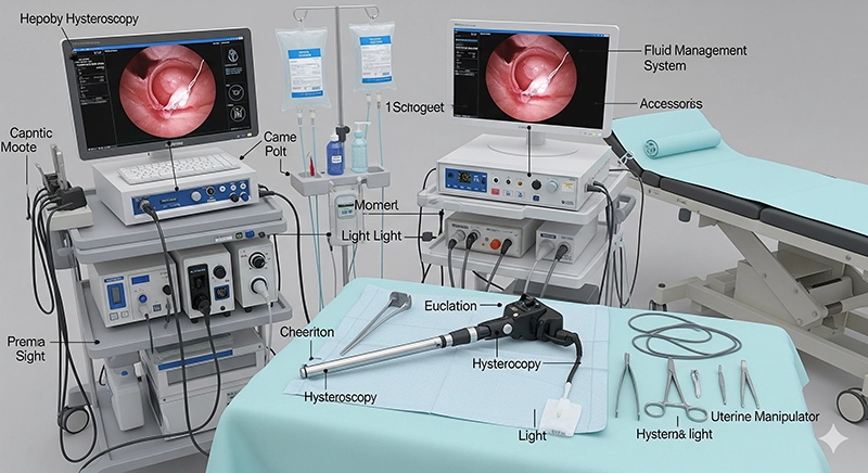
What makes the XBX system even more remarkable is its ecosystem approach. The hysteroscope can connect to other XBX imaging devices—such as the XBX video processor, LED light source, and recording system—to create a fully digital diagnostic chain. This connectivity ensures every pixel captured in the operating room becomes part of a permanent medical record.
Automatic color correction across all connected devices.
Simplified documentation for insurance and patient reports.
Real-time image transmission for telemedicine or consultation.
Centralized data storage, compliant with hospital information systems.
When all elements work together, surgeons no longer think about equipment—they think about results. That’s what integration means in the era of precision medicine.
A multicenter study conducted across five hospitals in Europe evaluated the XBX Hysteroscope’s performance in 500 patients. The outcomes were telling:
Overall diagnostic accuracy: 96%
Average operative time: 11.4 minutes
Complication rate: below 1%
Patient satisfaction: 98% rated “comfortable or very comfortable”
Numbers like these validate what surgeons have been reporting anecdotally for years. The XBX Hysteroscope doesn’t just detect uterine polyps—it redefines how gynecological precision should feel and function.
It leads to an important reflection: when evidence aligns with experience, that’s when technology truly earns its place in medicine.
So what’s next for XBX? The company’s R&D division is exploring AI-assisted pattern recognition that can automatically identify potential lesions and mark them on-screen for physician review. Imagine an interface that gently guides the surgeon’s eye, not replacing human judgment but enhancing it. Trials are already underway in cooperation with European teaching hospitals.
In addition, XBX engineers are testing lighter, wireless scopes with built-in processors—eliminating the need for bulky towers. These portable hysteroscopy solutions could soon make advanced diagnostics available even in smaller clinics or rural facilities.
In essence, the story of XBX hysteroscopy isn’t finished—it’s evolving with every patient, every image, and every surgeon who sees more clearly than before.
At its core, technology is only meaningful when it touches lives. A hysteroscope may seem like a tool of precision optics, but for the woman who finally understands her condition, it’s much more—it’s peace of mind.
When Mrs. Chen, a 39-year-old patient in Hong Kong, faced infertility after years of misdiagnosis, it was the XBX Hysteroscope that revealed a hidden polyp blocking implantation. After a minimally invasive removal, she conceived naturally within three months. Her doctor later said, “Sometimes it’s not about big surgeries; it’s about seeing what was once unseen.”
Stories like these remind us that medicine is not just science—it’s empathy illuminated through clarity.
Simplifying complexity—that’s the philosophy behind XBX. From high-resolution imaging to effortless fluid control, every detail of the hysteroscope reflects one goal: empowering doctors and comforting patients. The contrast between old and new isn’t just in pixels—it’s in outcomes, confidence, and dignity.
So yes, when we ask how XBX Hysteroscope detects and removes uterine polyps with precision, the answer is more than technical. It’s human. It’s about giving every woman the clarity she deserves and every clinician the confidence they need.
In the end, precision isn’t a promise. It’s a visible reality—one that shines every time an XBX lens enters the field of view.
The XBX Hysteroscope is designed for precise intrauterine visualization. It allows doctors to detect, diagnose, and remove uterine polyps or fibroids with minimal discomfort. Its high-definition optics and stable fluid management system provide clear, real-time images during hysteroscopic procedures.
Unlike traditional ultrasound or blind curettage, the XBX Hysteroscope provides direct visual access to the uterine cavity. Its integrated HD camera and adaptive illumination allow doctors to distinguish even small lesions, improving diagnostic accuracy and reducing false results.
Most patients experience only mild discomfort. The XBX Hysteroscope is designed with ergonomic size and smooth insertion tips to minimize irritation. Many procedures are done under local anesthesia or mild sedation, allowing same-day discharge and quick recovery.
Hospitals gain multiple advantages: reduced maintenance costs, shorter training time, and higher patient throughput. Because the XBX system supports both diagnostic and operative hysteroscopy, it helps medical teams complete procedures faster and more safely.

We not only provide sales of endoscope products, but also provide OEM/ODM customization services. We sincerely invite global partners to become industry innovators and share the hundreds of billions of market dividends. You are not only an agent, but also a strategic partner - your channel resources + our full-link empowerment = unlimited growth possibilities
Copyright © 2025.Geekvalue All rights reserved.Technical Support:TiaoQingCMS
