Table of Contents
The XBX medical endoscope is a precision imaging device designed to help physicians view internal organs and tissues with minimal invasiveness. It combines optical, electronic, and mechanical systems into a compact tool that provides real-time visuals of the body’s interior. Built under ISO 13485 and FDA-compliant standards, every XBX endoscope delivers stable performance, clear imaging, and safe operation during diagnostics and surgery.
A medical endoscope is a thin, flexible or rigid tube equipped with a camera, light source, and control handle that allows doctors to see inside the body without open surgery. The XBX medical endoscope transforms these functions into a unified platform that enables accurate diagnosis, biopsy collection, and treatment. For hospitals, this means faster recovery for patients, shorter operation times, and reduced infection risks.
Optical system: High-resolution lenses and image sensors capture bright, distortion-free visuals of internal cavities.
Illumination system: LED or fiber-optic light sources deliver consistent brightness for accurate visualization.
Control section: Ergonomically designed for precise handling, ensuring smooth navigation in narrow anatomical spaces.
Working channels: Enable suction, irrigation, and instrument passage during therapeutic procedures.
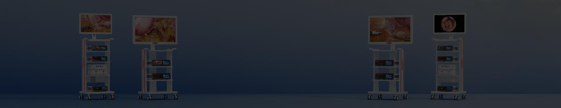
Unlike generic models, XBX medical endoscopes undergo rigorous testing for image fidelity, water tightness, and sterilization resilience. Hospitals trust XBX because of its consistent image performance, simplified maintenance, and compatibility across various endoscopy systems, including gastroenterology, urology, and ENT applications.
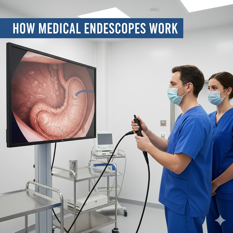
The XBX endoscope transmits light through a fiber bundle or LED at the distal tip, illuminating internal structures. The reflected light is captured by a CMOS or CCD sensor, converted into electrical signals, and displayed in real time on a medical-grade monitor. This visual feedback allows clinicians to diagnose abnormalities or perform treatments with minimal trauma.
The physician inserts the endoscope through a natural opening or small incision.
Light illuminates the internal organ, and the sensor sends video signals to the processor.
Images are enhanced by the XBX imaging system to highlight textures and blood vessels.
Doctors manipulate instruments through the working channel for biopsy, suction, or therapy.
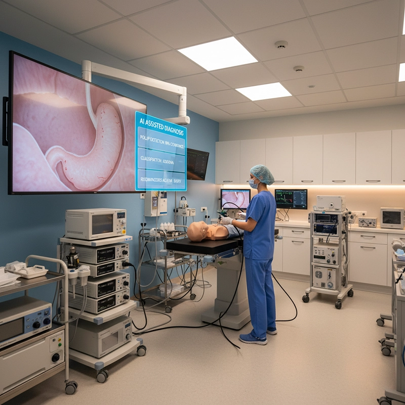
XBX uses advanced 4K and HD imaging technology with auto white balance and adaptive brightness control. The result is consistent color accuracy and tissue detail, even in deep or narrow regions where lighting is limited. The wide dynamic range preserves both bright and dark zones within the same field of view, which is crucial for precise surgical actions.
Video outputs are compatible with major operating room monitors and recording systems.
DICOM integration allows direct storage of images and videos into hospital archives.
Touchscreen interfaces simplify adjustments and data labeling during procedures.
 endoscopes and their uses
Endoscopes come in several specialized forms depending on the medical discipline. XBX produces a full range of endoscopic devices, each tailored for specific diagnostic and therapeutic tasks while sharing the same imaging core technology.
Flexible endoscopes: Used for gastrointestinal, bronchial, and urological procedures where access paths curve through anatomy.
Rigid endoscopes: Used for orthopedic, laparoscopic, and ENT surgeries requiring stable, straight pathways and high optical precision.
Gastrointestinal endoscopy: For viewing the esophagus, stomach, and colon to detect ulcers or tumors.
Bronchoscopy: For examining the airways and performing lung biopsies.
Hysteroscopy and laparoscopy: For minimally invasive gynecologic and abdominal surgeries.
ENT and urology: For diagnostic access to nasal passages, bladder, and urinary tract.
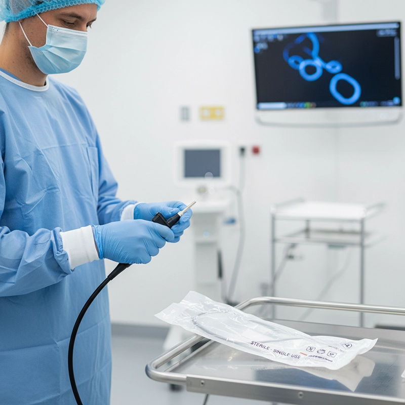
XBX manufactures both reusable and disposable models. Single-use endoscopes offer guaranteed sterility and eliminate reprocessing, while reusable models provide long-term value and durability. This dual offering allows hospitals to choose the right balance between cost and infection control.
Device longevity and patient safety depend on proper handling and sterilization. XBX medical endoscopes are constructed with sealed channels and chemical-resistant materials, reducing maintenance effort and minimizing downtime for clinical departments.
Leak testing is performed before cleaning to confirm device integrity.
Manual cleaning removes organic residue, followed by automated disinfection in an AER (Automated Endoscope Reprocessor).
Drying and visual inspection ensure the endoscope is ready for the next patient without cross-contamination risk.
Regular inspections verify articulation, image brightness, and channel patency.
XBX service teams provide calibration, spare parts, and firmware updates to maintain imaging accuracy.
Comprehensive documentation supports compliance with hospital quality systems and audits.
Hospitals choose XBX medical endoscopes for their balance of advanced imaging, ease of use, and clinical reliability. The combination of 4K visualization, robust materials, and global service networks gives healthcare providers confidence in both diagnostic and surgical performance.
Consistent imaging quality across specialties.
Certified safety and durability under ISO and FDA standards.
Flexible purchase options for reusable or disposable formats.
Comprehensive after-sales and training support.
The XBX medical endoscope represents a milestone in minimally invasive healthcare technology. By merging clarity, precision, and ease of integration, XBX continues to empower hospitals and surgeons worldwide to perform safer, faster, and more accurate procedures while maintaining patient comfort and clinical efficiency.
An XBX medical endoscope is a high-precision imaging device that allows doctors to observe internal organs and tissues in real time without open surgery. It combines a miniature camera, light source, and control system to transmit clear images from inside the body to a monitor during diagnostic or surgical procedures.
Light is delivered through fiber optics or LED illumination to the target area, and reflected light is captured by a high-resolution CMOS or CCD sensor. The signal is processed by an image processor, producing a live video feed on the surgical monitor, enabling physicians to detect and treat conditions accurately.
XBX medical endoscopes are used across multiple medical specialties, including gastroenterology (for colonoscopy and gastroscopy), pulmonology (for bronchoscopy), gynecology (for hysteroscopy), urology (for cystoscopy), and otolaryngology (for ENT examinations).
Both types are available. Reusable models are designed for long-term use and sterilization, while disposable endoscopes provide guaranteed sterility and eliminate the risk of cross-contamination—ideal for infection-sensitive departments like ICUs or emergency units.


We not only provide sales of endoscope products, but also provide OEM/ODM customization services. We sincerely invite global partners to become industry innovators and share the hundreds of billions of market dividends. You are not only an agent, but also a strategic partner - your channel resources + our full-link empowerment = unlimited growth possibilities
Copyright © 2025.Geekvalue All rights reserved.Technical Support:TiaoQingCMS
