
Table of Contents
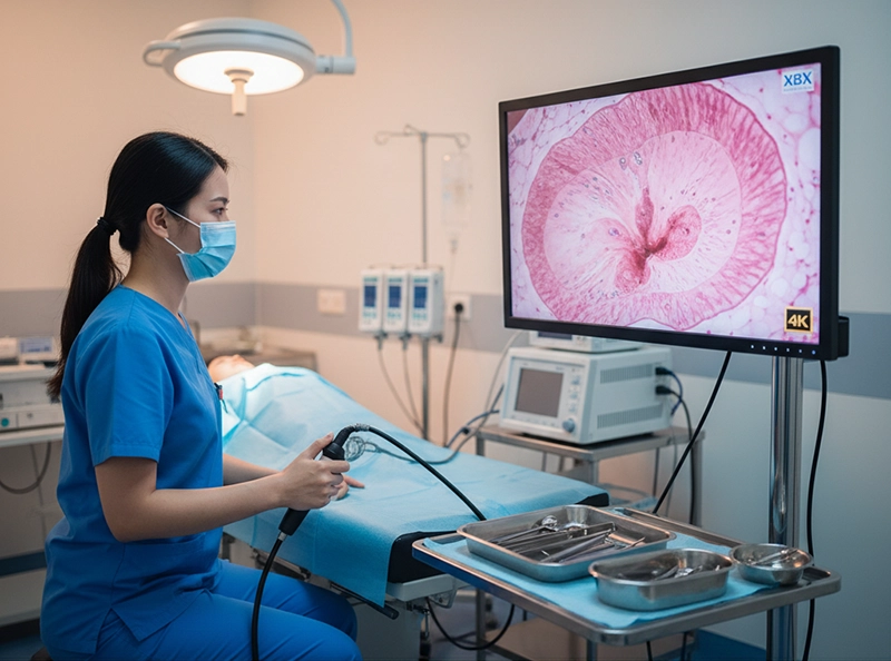
A hysteroscopy is a minimally invasive medical procedure that enables doctors to look directly inside the uterus using a thin, lighted instrument known as a hysteroscope. This scope, equipped with a camera and illumination system, is passed through the cervix into the uterine cavity, allowing real-time visualization on a monitor. Hysteroscopy is commonly used to investigate abnormal uterine bleeding, infertility, polyps, fibroids, adhesions, or structural anomalies. Compared with open surgery, it provides patients with faster recovery, less discomfort, and higher diagnostic precision.
Hysteroscopy answers the practical question of what is a hysteroscopy and what is hysteroscopy in daily medical practice: it is a direct, endoscopic view of the uterine cavity. By inserting a hysteroscope through the cervix, the gynecologist observes the endometrium in real time, records images, and, when indicated, performs treatment in the same session.
Hysteroscopy has transformed gynecology by offering direct visualization of the uterine cavity—something imaging techniques such as ultrasound or MRI cannot provide. It is now considered a cornerstone of modern women’s healthcare because it improves diagnostic accuracy, reduces unnecessary surgeries, and supports outpatient care pathways.
Improved diagnostic accuracy for small intrauterine abnormalities.
Dual role as both diagnostic and therapeutic tool in one encounter.
Patient-friendly, often completed in an outpatient setting with quick recovery.
Cost-efficient by reducing avoidable hospital stays and additional procedures.
Visualization: Ultrasound (indirect); MRI (cross-sectional); Hysteroscopy (direct uterine view)
Accuracy: Ultrasound (moderate for small lesions); MRI (high for large/complex lesions); Hysteroscopy (very high, even for small lesions)
Invasiveness: Ultrasound (non-invasive); MRI (non-invasive); Hysteroscopy (minimally invasive)
Treatment Ability: Ultrasound (no); MRI (no); Hysteroscopy (yes: diagnosis + treatment)
Hysteroscopy can reveal and treat a wide spectrum of intrauterine conditions by allowing the clinician to see and address the problem at its source.
Abnormal uterine bleeding: Heavy, irregular, intermenstrual, or post-menopausal bleeding can be investigated to identify structural causes or endometrial changes.
Endometrial polyps: Benign overgrowths of the lining that may contribute to bleeding or infertility; hysteroscopy enables direct visualization and removal.
Submucosal fibroids: Fibroids protruding into the cavity often cause heavy bleeding and fertility issues; hysteroscopic resection precisely targets the lesion.
Uterine adhesions (Asherman’s syndrome): Scar tissue that can distort the cavity, leading to infertility or altered cycles; adhesiolysis restores normal anatomy.
Congenital uterine anomalies: Septum or other variants may impair fertility; hysteroscopy confirms and sometimes corrects these anomalies.
Suspected hyperplasia or malignancy: Targeted, direct-vision biopsy improves diagnostic yield for premalignant or malignant lesions.
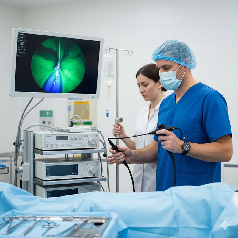
The procedure follows standardized steps prioritizing safety, comfort, and clear visualization.
Individualized anesthesia plan (none, local, or general depending on complexity).
Cervical preparation or gentle dilation if needed.
Preparation of distension media (saline or CO₂) to open the uterine cavity for viewing.
The hysteroscope passes through the cervix into the uterine cavity under direct vision.
Saline or CO₂ gently expands the cavity to improve visibility.
The endometrium is inspected systematically; images are recorded for documentation.
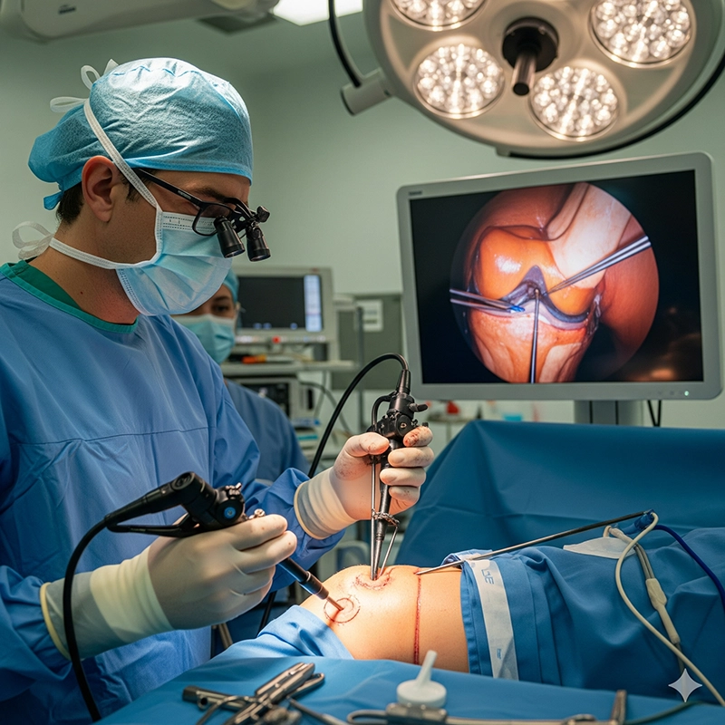
When indicated, miniature operative instruments are introduced to treat pathology.
Most patients go home the same day and resume activities within 24–48 hours.
Mild cramping or light bleeding can occur temporarily.
Follow-up is scheduled to review findings and next steps.
Purpose: Diagnostic (observation); Operative (diagnosis + treatment)
Duration: Diagnostic (about 10–15 minutes); Operative (about 30–60 minutes)
Equipment: Diagnostic (basic hysteroscope); Operative (hysteroscope + surgical instruments)
Outcome: Diagnostic (visual confirmation/biopsy); Operative (removal/correction/biopsy)
Hysteroscopy balances high diagnostic yield with minimal invasiveness, making it a widely adopted option in modern gynecology.
Combines diagnosis and treatment in one session when clinically appropriate.
Faster recovery and reduced post-procedure discomfort compared with open surgery.
Fertility-preserving where feasible by targeting intrauterine pathology precisely.
Often performed as an outpatient procedure, supporting efficient care pathways.
Infection requiring observation or antibiotics.
Uterine perforation (uncommon, managed per clinical protocols).
Unexpected bleeding; most cases are self-limited.
Reactions associated with anesthesia when used.
In fertility care, hysteroscopy plays a central role by ensuring the uterine cavity is receptive to implantation. Before IVF, many clinics assess and, if necessary, optimize the cavity. In recurrent miscarriage or unexplained infertility, hysteroscopy identifies correctable lesions such as polyps, adhesions, or septa, helping align the uterine environment with reproductive goals.
Use of hysteroscopy continues to expand globally as awareness of women’s health grows and minimally invasive techniques become standard. Technological advances enhance image quality and workflow while broadening access to care in outpatient and resource-limited settings.
Disposable hysteroscopy equipment to streamline reprocessing and minimize cross-contamination risk.
4K/HD visualization that improves tissue differentiation and clinical confidence.
AI-assisted pattern recognition supporting early detection and documentation consistency.
Portable hysteroscopy machines that extend services to clinics outside major centers.
Beyond the clinical lens, understanding the ecosystem surrounding devices helps hospitals and clinics align technology choices with safety, training, and sustainability. This section introduces essential B-side concepts while keeping a science-popularization tone.
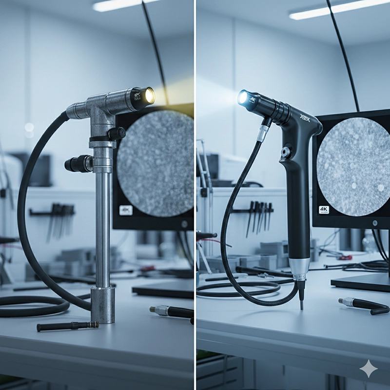
Core components: hysteroscope (rigid or flexible), camera/monitor, LED or xenon light source, distension media unit, miniature operative instruments.
Clinical impact: reliable optics and stable fluid management enhance safety and visualization.
Maintenance: routine checks, proper reprocessing, and staff training sustain performance.
Integrated systems combine visualization, illumination, fluid control, and instrument channels.
Modern designs emphasize ergonomics, digital recording, and EMR connectivity.
Compact/portable models support office-based procedures and outreach clinics.
Production under ISO 13485 with medical-grade materials and validated sterile workflows.
Precision optics and assembly lines ensure consistency and device reliability.
R&D collaborations with clinicians translate feedback into safer, more effective devices.
Selection factors: certification portfolio (CE/FDA/ISO), breadth of diagnostic/operative systems, after-sales training and support.
OEM/ODM options help hospitals match instruments to specialty workflows and budgets.
Lifecycle support covers spare parts, upgrades, and user education.
Role: connect factories/manufacturers to hospitals, manage logistics, installation, and local training.
Value: timely access to upgrades, consumables, and technical assistance that keeps services running smoothly.
Example: XBX provides endoscopy-focused supply solutions pairing advanced hysteroscopy equipment with training programs and long-term service support, helping procurement teams balance technology, safety, and continuity.
Hysteroscopy is a bridge between accurate medicine and minimally invasive care. For patients, it offers a safe, effective approach to diagnosing and treating intrauterine conditions. For clinicians, it delivers precision and efficiency. For healthcare organizations, it is a strategic investment. And across the industry, continuous innovation in hysteroscopy equipment, integrated hysteroscopy machines, quality-driven hysteroscopy factories, responsible hysteroscopy manufacturers, and reliable hysteroscopy suppliers—such as XBX—collectively advances women’s health.
XBX offers both diagnostic and operative hysteroscopy systems, including high-definition imaging scopes, ergonomic instruments, and complete fluid management setups suitable for gynecological care.
Yes, XBX provides OEM and ODM options, allowing hospitals to adapt hysteroscopy equipment to their clinical protocols, budgets, and space requirements.
XBX products comply with international medical device standards, ensuring compatibility with hospital procurement processes in multiple global regions.
XBX hysteroscopy systems integrate fluid control technologies, high-quality optics, and precise operative tools to minimize risks such as fluid overload, infection, or uterine perforation.
Yes, XBX offers slim, flexible scopes designed for office-based hysteroscopy, enabling hospitals to expand minimally invasive services without the need for full operating theaters.
XBX supports distributors with OEM/ODM branding, competitive pricing, flexible order volumes, and strong after-sales backing, ensuring market growth opportunities.
XBX focuses on miniaturized scopes, ergonomic designs, and advanced imaging to make outpatient hysteroscopy more accessible, aligning with global gynecology trends.
Hysteroscopy is a minimally invasive procedure where a thin scope is passed through the cervix into the uterus to diagnose or treat intrauterine conditions.
Hysteroscopy is used to detect polyps, fibroids, adhesions, septa, hyperplasia, and suspected endometrial cancer.
Diagnostic hysteroscopy visualizes the uterine cavity, while operative hysteroscopy includes instruments to treat pathologies during the same session.

We not only provide sales of endoscope products, but also provide OEM/ODM customization services. We sincerely invite global partners to become industry innovators and share the hundreds of billions of market dividends. You are not only an agent, but also a strategic partner - your channel resources + our full-link empowerment = unlimited growth possibilities
Copyright © 2025.Geekvalue All rights reserved.Technical Support:TiaoQingCMS
